Photometry
Photons enter the eye through the optic lens, which, in the human eye, is controlled by muscles which flex it in order to focus the light on different portions of the retina, which extracts salient features from the environment and encodes them in the spike trains of retinal ganglion cells, which are the output neurons of the eye. There are over 40 ganglion cell types. Amongst the matter absorbing these photons are photoreceptive cells, rods, and cone cells. These cells are preferentially sensitive to light of varying wavelengths and intensity. The absorption of photonic energy catalyzes a reaction in a receptive protein molecule, of class known as opsins, creating a chain reaction of protein activity for the purposes of cell signaling. The degree and duration of this stimulus either excites or inhibits the cell, towards a threshold where the cell will be more or less inclined to fire, sending a bioelectrical signal to another kind of specialized nerve cell, as the first step of relaying the signal that carries information as to what has happened with that particular receptor into the optic nerve and ultimately into the brain, where the combined stimulus of all the receptors is amalgamated, filtered, and processed to create visual perception and the subjective experience of vision.
Rod phototransduction drives the pupillary contrast response. These are 3 classes of photoreceptors in the mammalian retina: rods, cones, and intrinsically photosensitive retinal ganglion cells. Approximately 150 different types of neurons are connected in about 45 distinct circuits (40 circuits as of 2020). These circuits convert the pixel representation of photoreceptors into parallel feature representations of ganglion cells.
Rod cells and cone cells.
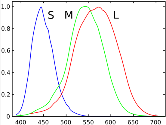
Photoreceptor cells convert light (visible electromagnetic radiation) into signals that can stimulate biological processes. Cone cells are photoreceptor cells in the retinas of vertebrate eyes and are responsible for color vision, functioning in bright light, whereas rod cells, work better in dim light. Rod cells have little role in color vision, which is the main reason why colors are much less apparent in dim light. There are 3 types of cone cells: S-cones, M-cones, and L-cones, for short, medium, and long. When violet light strikes the eye for example, the S-cone is stimulated strongly, and the L-cone stimulated weakly.
There’s about 92 million rod cells in the human retina, and 4.5 million cone cells in the human eye.
Humans having red green and blue cones, instead of red green and violet cones, explains why in the Doppler effect, we call it blue shift instead of violet shift. Our red and green cones have a closer overlap, meaning we see yellow better because it affects both our red and green cones.
Now, there is a misconception. Our red cones are not actually red, they're peak is in the yellow range. The halfway point between red and green in the spectrum, is yellow, and if you Google'd what color do humans see the best, majority return green. But you can find a few, that actually say chartreuse, or whatever color is 555 nm. That is roughly 50% green and 50% yellow, so our red cones are mistakenly labeled red. This makes sense, as we cannot see in the IR. Unfortunately, the decision to call it red cones was already established by 1931, and it could have been due to marketing reasons.
Therefore, how does humans having yellow cones instead of red, leave humans with red-green colorblindness? Here's the simple answer prior to 2023: has to do with opponent processing. Even though we have 3 cones, we see 4 colors. The opposite color, is when we stare at a color for long, and close our eyes, we see the opposite color. For blue and yellow, are themselves opposite. And for green, is red.
However, a paper by Conway, Malik-Moraleda, and Gibson published in July 2023, went to disprove Hering's opponent-colors theory (1878). It did not disprove Pridmore's 2013 paper on complementary colors (red-cyan, green-magenta, and blue-yellow).
After July 2023, the new version is: color vision begins with 3 types of cones, but our perception of color results from complex neural processing beyond the cones. Color adaptation can produce complementary afterimages (staring at red can produce a cyan afterimage, green can produce magenta, and blue can produce yellow, and vice versa).
Pridmore's 2013 paper was attacking an important physiological pillar of Hering's theory, whereas Conway et al. went further and argued against the entire Hering framework for explaining color appearance.
Red-green and blue-yellow colorblindness.
8% of men are red-green colorblind. But among those 8%, have some variances.
Anomalous trichromacy: ~6% (mild red-green colorblindness).
Protanomaly (L cone shifted towards M): ~1%
Deuteranomaly (M cone shifted towards L): ~5% (most common)
Dichromacy: ~2% (severe red-green colorblindness).
Protanopia (no L cone function): ~1%
Deuteranopia (no M cone function): ~1%
Tritanopia (no S cone function): 1 in 100,000
Protanopia is a severe form of red-green color blindness, in which the L-cone is absent. It affects about 1% of males. Colors of confusion include blue/purple and green/yellow. Red appears darker.
Deuteranopia is a severe form of red-green color blindness, in which the M-cone is absent. It affects about 1% of males. Still colors of confusion between blue/purple and green/yellow, but red does not appear darker.
Tritanopia is a severe form of blue-yellow color blindness, in which the S-cone is absent. They tend to confuse greens and blues. It is much rarer than the other types, occurring in about 1 in 100,000, but is not sex-linked, so affects females and males at similar rates. Tritanopia and tritanomaly are both autosomal. L and M opsin genes are on the X chromosome, while the S-cone opsin gene (OPN1SW) is on chromosome 7. The 2 red-green colorblind genes, OPN1LW and OPN1MW are both on the X chromosome.
Now, this should sound like a contradiction for protanopia, as we called it red-green color blindness, but is actually yellow-cones. Well, even though it peaks in the yellow range, color is encoded by comparing cones, not by where they peak. So yellow is computed with L + M cones together, with little S. Blue-yellow colorblind means the person can still see yellow.
Normal:
Red–Green channel = L - M
Blue–Yellow channel = S - (L + M)
In protanopia:
L = 0
So Red–Green channel = 0 - M = -M
Blue–Yellow channel = S - (L + M) = S - M
The cleanest way to think about it is instead of "we have blue, green, and yellow cones," we have 3 types of cones with different spectral sensitivity curves. The visual system combines their signals, including differences and sums, to extract information about wavelength and perception.
Now for protans (1% + 1%), they can generally see green better than red. Because they lake the yellow cones, the boundary of where normal humans can no longer see red into IR, is shifted. And green appears more yellow.
Women can be red-green colorblind: .5% so 1 in 200.
Women have 2 X chromosomes, but most cells don't normally use both X chromosomes simultaneously. Early in embryonic development, each cell essentially chooses 1 X chromosome to keep active and largely shuts down the other (called X-inactivation). Since men are XY, they only have 1 X chromosome, and if his X chromosome carried a defective L or M opsin gene, he has no 2nd X chromosome carrying a normal copy, to compensate. If a woman has 1 X carrying the variant and the other X carrying a normal version, she is often a carrier without the colorblindness. So, women having 2 X chromosomes means 8% * 8% is .64%, though in medical literature, .5% is used as the % of women that are red-green colorblind.
-
Rhodopsin is a protein that is the opsin of rod cells in the retina. Rhodopsin reactions can take 50 femtoseconds.
The 6 types of photoreceptor proteins are: cryptochromes, phototropins, BLUF proteins, phytochromes, xanthopsins, and rhodopsins. Only the 1st 3 bind to flavins, while the last 3 use other chromophores to absorb light. Flavins are always yellow in their oxidized form (not reduced). However, only the 1st and 6th are found in animals.
Cryptochromes are found in animals, plants, and bacteria, and regulate circadian rhythm. Phototropins are primarily found in plants and algae. BLUF proteins (blue light using FAD) are found in bacteria and some algae. Phytochromes are found in plants, fungi, and bacteria, and bind to a chromophore called bilin (usually phytochromobilin). Xanthopsins are primarily found in bacteria. Rhodopsins are found in animals, fungi, and bacteria.
Reflective light.
Most materials have some absorption cutoff where shorter-wavelength radiation cannot penetrate, because it moves an electron across a band gap. Metals tend to reflect radiation from radio waves to UV. But colored metals like copper and gold don’t reflect the shorter wavelength (Webb space telescope cannot see blue or UV reflected from gold).
In 2020, 3-hydroxyisonicotinaldehyde (also known as 3-hydroxypyridine-4-carboxaldehyde), has the lowest molecular weight of all dyes, which exhibits green fluorescence.
Do animals see color?
Animals such as rabbits and alligators, lack the red cone, but do have the blue and green cone. That generally means they can see blue and green the same way we do, but not red.
Most birds see orange through ultraviolet. Few birds can see into the red range of the spectrum. Other animals like bumblebees cannot see into the red spectrum. Turtles, however, see an excess of red, and they can also see ultraviolet. It is a myth that goldfish can see from ultraviolet to infrared, and it turns out no known animal can. And the birds and turtles that see into ultraviolet, see into UV-A, not UV-B.
-
1. If animals such as rabbits and alligators, lack the yellow cone, but do have the blue and green cone. Then do any animals lack the green cone, but do have the yellow and blue cone?
The problem with humans, is that out S (short) and M (medium) cones are so close aligned, that when an animal does have only 2 cones, it's hard to say whether the shorter cone, is for S or M. Note that for humans, the red cone is mistakenly called red. The red (or S) cone, is actually peaked in the yellow region. The idea of calling them blue, green, and red cones, instead of blue, green, and yellow cones, was already decided by 1931, and may have been due to marketing reasons.
While no common animal species are documented to naturally possess only yellow and blue cones, it’s theoretically possible through genetic mutations.
Most animals that have only 2 cones, are in the blue, and yellow-green region, such as freshwater fish. Note that orange or red cones is especially unuseful in underwater environments, as red light are often filtered out by water.
2. What about lacking the blue cone, but have the yellow and green cone?
Still no.
3. What animals have only 1 cone, and what color does it fall under?
Seals and whales, and it's believed they've lost the blue cone from evolution, and the only other cone they have is for seeing green. Whales and dolphins only see into the yellow-green region, with seals, sea lions, and walruses only seeing into the green region. In deep water, having a cone sensitive to green-yellow helps with contrast detections against a blue background.
3. Any animals with other combinations?
Dogs and wolves do not see red or green, and only see blue and yellow-green (and therefore brown and gray). Dogs have 2 types of cone cells in their retinas: blue sensitive cones (L-cones) and yellow-green sensitive cones (429 and 555 nm). Because dogs cannot see red, then they see purple without the red, so purple looks blue to them.
Note: it should be noted that most people believed dogs were completely colorblind, especially due to optometrist Gordon Walls publishing a book in 1942, "The Vertebrate Eye and Its Adaptive Radiation." But that belief was debunked by Aug. 1989, when ophthalmologist Dr. Jay Neitz, then at the University of California, Santa Barbara, and his colleagues Timothy Geist and Gerald Jacobs discovered that canines could see blues and yellows but not reds and greens. They discovere dogs are dichromats, compared to humans being trichromats.
The lab mouse, has a cyan-cone and ultraviolet cone (508 nm and 360 nm).
4. What about cats? Did Walls also believe cats were completely colorblind? Who was the 1st to prove cats can see some colors?
Cats are at 454 and 561 nm, according to a Jan. 1987 paper. But a April 1993 paper argues 450 and 550 nm. Walls did believe cats were completely colorblind as well. However, a later thesis summarizing Walls' 1953 discussion reports that Walls acknowledged color-sensitive units in the cat retina, but suggested that their physiological significance might not be related to the animal's color vision. This was in the context of work finding wavelength-dependent responses in cat retinal fibers.
But who 1st showed that cats can actually see color? The answer appears to be Daw & Pearlman, 1970. N. W. Daw & A. L. Pearlman (1970), “Cat colour vision: evidence for more than one cone process,” Journal of Physiology 211:125–137.
They trained living cats to distinguish red from cyan, and orange from cyan. They tested the animals under conditions in which the rods should have been saturated. The cats continued to discriminate the colors, and one cat could still distinguish red from cyan after exposure to extremely bright white light.
It should be noted that in 1977, a paper wrongly claimed cats were trichromats. Titled “Trichromatic vision in the cat” reported 3 cone systems, by Ringo, Wolbarsht, Wagner, Crocker & Amthor, suggesting 450, 500, and 555 nm. That was later disproved by the Jan. 1987 article "Photopic spectral sensitivity of the cat" by M. S. Loop, C. L. Millican, and S. R. Thomas. Then came the April 1993 paper, Guenther & Zrenner "The spectral sensitivity of dark- and light-adapted cat retinal ganglion cells."
Here is where specifically Walls called cats colorblind in his 1942 book: on page 338 of The Vertebrate Eye and Its Adaptive Radiation, in the discussion of the lateral geniculate nucleus, Walls writes “And, the cat, which has no color vision, has almost as complex a geniculate as man…”
5. What other animals have 3 cones?
Besides the primates, animals that are like humans and have 3 cones include many fish (goldfish) and lizards.
6. Which animals have 4 or more cones?
Most birds have 4 cones, allowing them to see more into the UV. The Mantis shrimp holds the record at 16 cones.
7. If animals have 1 cone, then how can some animals be completely colorblind?
The difference is cone monochromacy, and rod monochromacy. For the 2nd, those animals have retinas that have rods, with no functional cone system. Examples of these include some sloths, armadillos, and golden moles.
8. What about rod dichromats or rod trichromats?
Still none.
9. Who was the 1st animal to be discovered to see color?
In 2014, a modern review calls honeybees the 1st animal in which color vision was convincingly demonstrated. Lubbock had already reported in 1882 that honeybees repeatedly visited colored cards when rewarded with honey. But the much more convincing demonstration came from von Frisch in 1914. Von Frisch 1st trained bees to a coloured card by rewarding them with sucrose solution. He also discovered that honeybees were red-blind. The honeybee eye contains 3 types of photoreceptors which peak in the UV, blue, and green parts of the spectrum.
10. When was the so-called red cone actually discovered to be in the yellow region?
When were the 3 human cone peaks actually measured? 1964. 2 groups independently used microspectrophotometry to measure the absorption spectra of individual human cone photopigments:
Brown & Wald (1964).
Marks, Dobelle & MacNichol (1964).
Before that, the Young–Helmholtz theory was commonly described as 3 retinal mechanisms producing sensations of red, green, and violet/blue. For example, the 1911 Encyclopædia Britannica describes Young's 3 as red, green and violet, while noting Maxwell's evidence favored red, green and blue.
11. What happens when you mix red and blue?
It depends on whether you meant paint or light. For paint, then purple, and for light, then magenta.
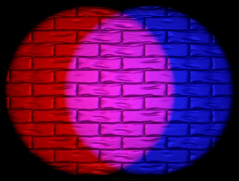
The following is an image off 500 red and blue pixels side by side. You can try to download it, then zoom-in to see the distinct pixels.

Now, as far as making side by side pixels of other colors, keep in mind that your computer or smartphone screen uses red green and blue pixels, so if I used other colors, are not represented by the light of your screen.
12. What happens when you mix red and green?
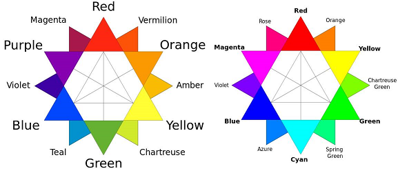
It also depends on what you mean by green. For red and blue, they are essentially the same between the paint-model of colors, and light-model of colors.
The paint model shows a darker green, whereas the light model shows a lighter, lime green.
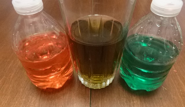
Therefore, the word green is often used to represent 2 different colors. Below is an example of mixing red and green liquid.
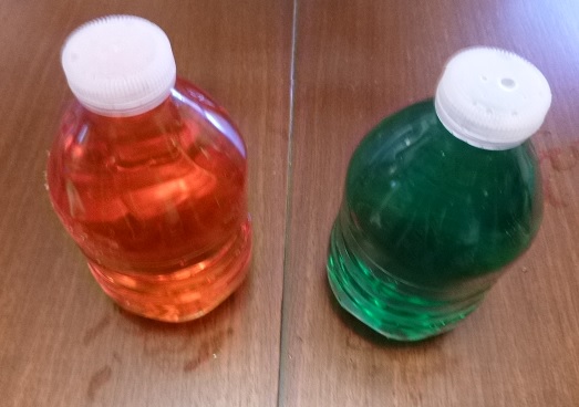
However, unfortunately, the red liquid is not that red to me. It is a bit to the orange side. Another dye I bought, was more orange than this.