Human Anatomy
Cells.
The human body contains around 37 trillion cells. Bacteria outnumber eukaryotic cells in our body about 10 to 1.
The average skin cell has a diameter of 30 micrometers. Every hour your body creates 20 million new skin cells, which push older cells up in the epidermis. Every square cm of skin is shedding about a 1,000 dead skin cells/hour. That’s about a lb of dead skin in a year.
Blood.
By volume, blood is about 55% liquid and 45% cells. The liquid portion of blood is called plasma. All blood cells are formed from hematopoietic stem cells, which originate in the bone marrow.
Each red blood cell contains about 250 million molecules of hemoglobin, each of which can bind 4 molecules of oxygen. Normal amounts of hemoglobin in the blood for males is 13.5 to 17.5 g/dL, 12.0 to 16.0 g/dL for women. Normal hematocrit (the % of red blood cells in the blood sample) for males is 41-53% and 36-46% for women.
White blood cells (leukocytes) usually comprise <1% of total blood volume, which is about 4,500 to 11,000 leukocytes per microliter of blood. About 60% of white blood cells are neutrophils, 30% are lymphocytes, and about 8% are monocytes. There are 2 types of lymphocytes: B-lymphocytes and T-lymphocytes (B-cells and T-cells). B-cells produce proteins as antibodies, called immunoglobulins (Igs), while T-cells do the attacking.
Glucose makes up about .10% by mass of human blood. Approximately 4 g of glucose are present in blood at all times, and about 18 g total in the human body. The adult human liver produces 180-220 g of glucose in 24 hours.
The entire volume of a person’s blood is filtered by the kidneys about every 40 minutes.
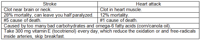
Difference between strokes and heart attacks:
Coronary artery bypass graft (CABG) surgery:
-About 200,000 Americans have it each year, with 40-50% being over 65.
-Average cost of $46,800, so $9.3 billion spent a year.
-Takes 3-6 hours, performed by cardiac surgeons whom are private practice, and operate at multiple hospitals.
CABG surgery is to treat coronary artery disease, which is the buildup of plaques in the arteries of the heart. There is no equivalent for veins in the heart, as that is uncommon as veins have lower blood pressure.
Hemostatis agents.
Following the Sept. 11, 2001 attacks, the U.S. military was looking for new technologies to stop arterial bleeding. Dr. Alan Hassan of Northwestern University, was the 1st to come up with the best known hemostatis agents, as well as high-tech medical products. The winnter was something called QuikClot, a grainy Ca 5A zeolite. It is a powder that can be dumped onto gushing wounds to stanch arterial bleeding, packaged as Z-Medica's 1st product. It originally had a high release of heat problem, and the Navy went to Professor Stucky from UC Santa Barbara for help. The zeolite had released so much heat, that led to the body's platelet activation. In 2013, the FDA approved of QuikClot Combat Gauze for topical surgical wound and traumatic bleeding.
Biological chemistry:
O2 and CO
This will attempt to explain what it is about oxygen that keeps us alive. It is a slight misconception to say oxygen keeps the brain alive. It does not do so directly. ATP keeps the brain alive, and so oxygen itself does not directly keep the brain alive.
1. What is it about oxygen (O2) that keeps us alive?
There are 2 broad answers to this, 1 is oxygen is used in combustion reactions in our body (catabolism), and the other is oxidative phosphorylation chains (cellular respiration), where O2 is ultimately reduced to water.
When we’re not jogging, 85% of the oxygen is used in cellular respiration, and 15% used in catabolism. When we’re jogging, the ratio is more 90/10. Also, < 1 % of oxygen is used for scavengering free radicals.
Oxygen is the final electron acceptor in the electron transport chains in cellular respiration. Its purpose is to produce ATP. As a final electron acceptor, it is responsible for removing electrons from the system. If oxygen were not available, electrons could not be passed among coenzymes, the energy in electrons could not be released, the proton pump could not be established, and ATP could not be produced.
Ultimately, oxygen receives 2 electrons and 2 protons to form water. Oxygen does not receive electrons and protons as O2 as some textbooks say, but only as a single oxygen atom. If it were O2, it would form hydrogen peroxide (H2O2), which is more acidic and an oxidizer.
2. What about carbon monoxide, CO?
Carbon monoxide also has an oxygen atom, so why can’t it accept as the final electron acceptor in the transport chain? Carbon wants 4 bonds, so in a C=O, carbon has a lone pair. Unlike oxygen, carbon monoxide does not bind off hemoglobin. Carbon monoxide binds to hemoglobin at a rate of 210 times more than oxygen. So the oxygen that does not get binded to hemoglobin does not continue its rule in ATP production.
The human brain generally can last 3 minutes without ATP, however, some parts of the brain can only last 90 seconds without ATP.
You may see in many texts that the preferred fuel for the brain is glucose, which obviously can cross the blood-brain for energy for the brain. (Brain tissue does not store energy.). So the brain relies on circulation for its fuel supply. However, it is ultimately ATP that is fuel for the brain. Glucose and ketones make ATP in the brain. Brain tissue can also adapt to ketone bodies such as acetoacetate as a source of fuel.
The triple bond in carbon monoxide is also the strongest known bond in chemistry for a stable molecule (things like steel however, are not a molecule).
H2O
3. What is it about water that keeps us alive?
Water is the medium in which a lot of reactions take place in the body. Without water, those reactions wouldn’t happen.
There is no substitute for this. The nitrogen-equivalent for water is ammonia (NH3). NH3 exists as a gas in room temperature and in the body, so it cannot be a substitute. Going down the column, H2S is also a gas. Hydrogen peroxide (HOOH) is slightly more acidic than water, and so the pH is off.
However, not all reactions in the body take place in water, such as reactions within membranes. Besides being a medium for reactions, water also organizes the shape of proteins.
The autonomic nervous system.
The nervous system is divided into 2 anatomical division: the central nervous system (CNS), which is composed of the brain and spinal cord, and the peripheral nervous system, which includes neurons located outside the brain and spinal cord (that is, any nerve that enters or leaves the CNS). The peripheral nervous system is further divided into 2 parts: the efferent division (the neurons that bring information from the brain and spinal cord to the peripheral tissues), and theafferent division (the neutrons that bring information from the periphery to the CNS). The efferent division is further divided into 2 parts, the somatic (the voluntary control of skeletal muscles) and autonomic systems (the vital body functions without conscious participation of the mind).
The most common central nervous system tumor is meningioma, then glioblastoma.
Immunology.
There are 5 classes of immunoglobulins: IgG, IgM, IgA, IgD, and IgE. Each class of immunoglobulin has different functions and may be more effective against certain types of bacteria. All of them are produced by B cells. Specifically, all of them are produced by plasma cells, which are the activated form of B cells.
IgG, is the most abundant immunoglobulin in the blood and tissues, and is capable of neutralizing a wide range of both gram-positive and gram-negative bacteria, and pathogens. IgM is the 1st immunoglobulin produced during an infection and is effective in activating other components of the immune system to fight bacteria. IgA is primarily found in mucosal areas, such as the respiratory, gastrointestinal tracts, saliva, and tears, and provides protection against bacterial invasion at these surfaces. IgD is present in small amounts in the bloodstream, and is believed to play a role in the activation of B cells (a type of immune cell that produces antibodies) and is typically found on the surface of B cells. IgE is involved in the immune response against parasitic infections and plays a central role in allergic reactions. When a person is exposed to an allergen (e.g., pollen or pet dander), IgE antibodies trigger the release of inflammatory substances, such as histamine, leading to allergy symptoms.
Immunotherapy works by stimulating the body’s natural defense system to attack cancer.
Existing antibody drugs used in cancer belong to an antibody type called IgG, but as of summer 2023, IgE antibodies have not been tested in humans before. IgE antibodies evolved to target parasites like worms and flukes, and IgG antibodies are involved in attacking bacteria and viruses in the body. IgE is more commonly known as the antibody associated with allergic reactions. If you’ve ever had an allergy blood test, this is what the doctor will be looking for.
Cytokines are how cells illicit an immune response, and an immune response activates all sorts of things that bring stress on cells, such as reactive oxygen species. Besides infectious agents, allergens, and cancer, literally anything that affects capillary permeability or causes tissue damage, will cause the body to create more antibodies.
Interleukins.
1 such group of cytokines are interleukins, which are proteins and signal molecules secreted by white blood cells (as well as other body cells). The human genome encodes more than 50 interleukins. The function of the immune system primarily depends on interleukins, and the majority of interleukins are synthesized by CD4 helper T-lymphocytes and hematopoietic cells. There is some evidence that interleukin-17 makes worms and mice more social. There's a lot of theories on how interleukin-17 crosses the blood-brain barrier. Interleukin-25, discovered in 2001, is also known as interleukin-17E.
Other cytokines besides interleukins, include chemokines, interferons, and lymphokines.
Short term vs. long term allergies.
Immediate hypersensitivity (like hives and swelling after eating lobster in someone with shellfish allergy) occurs rapidly because the immune cells reacting had already been sensitized (they are bound by IgE antibody that recognizes the allergenic protein in shellfish). Therefore, exposure to the allergen signals through IgE binding and those cells release pre-formed allergy mediators (histamine mostly). Delayed type hypersensitivity (like being allergic to poison ivy and having rash peak at 1-2 days after exposure) is due to a direct cellular immune response. T-cells that recognize the allergen in poison ivy become activated and have to secrete chemokines and other inflammatory signals that result in recruitment of inflammatory cells and this inherently takes more time.
Diabetes.
As of 2012, diabetes hits an estimated 23.6 million in the U.S., and over 250 million worldwide. As of 2015, to 25.8 million in the U.S., and 347 million people worldwide. The American Diabetes Association recognizes 4 types of diabetes: type 1 (formerly called insulin-dependent diabetes mellitus), type 2 (formerly non-non-insulin-dependent diabetes mellitus), gestational diabetes, and diabetes due to other causes such as genetic defects or medications. Type 2 accounts for more than 90% of cases.
The pancreas produces the peptide hormones insulin, glucagon, and somatostatin, which are secreted from the cells in the islets of Langerhans. A lack of insulin, as seen in diabetes mellitus, can cause serious hyperglycemia.
Neurobiology.
Memory is believed to be stored mainly in the pattern and strength of synaptic connections between neurons, in the network architecture and synaptic weights, mostly in dendrites and their spines, not inside the neuron like files in a folder.
The brain is the most energy-efficient computational device yet encountered, requiring only about 20 Watts of power to sustain the physical processes that govern brain function, including thought and consciousness. For a baby, can have a mom that speaks 1 language, a dad that speaks a 2nd language, and an at-home maid that speaks a 3rd language, and by age 4, the toddler can translate the languages, all while their food intake is about 1400 kilocalories a day, or about 70 Watts, in which more than half is used by their brain.
Microglia are a type of neuroglia (glial cell) located throughout the brain and spinal cord. Microglia account for about 10-15% of cells found within the brain. As the resident macrophage cells, they act as the first and main form of active immune defense in the central nervous system (CNS).
Rubicon is a protein that in humans is encoded by the RUBCN gene, and is consisted of 972 amino acids. It is 1 of the few known negative regulators of autophagy, a cellular process that degrades unnecessary or damaged cellular components. Rubicon is recruited to its sites of action through interaction with the small GTPase Rab7, and impairs the autophagosome-lysosome fusion step of autophagy through inhibition of PI3KC3-C2 (class III phosphatidylinositol 3-kinase complex 2).
The ventral pallidum is part of the basal ganglia and plays a key role in reward, motivation, and addiction. In the 1960s and 1970s, neuroscientists recognized it as part of the "limbic loop" within the basal ganglia, distinct from the dorsal pallidum (which includes the globus pallidus) and associated with reward pathways.
The nucleus accumbens is also part of the basal ganglia and critical in reward, pleasure, and reinforcement learning. By the 1970s and 1980s, the nucleus accumbens was increasingly studied for its role in reward circuitry, and researchers identified it as a major site of action for neurotransmitters like dopamine in processes related to addiction and motivation.
ARCH vs. eYFP.
In neurobiology, ARCH and eYFP refer to tools used in optogenetics and imaging studies to manipulate or observe neuronal activity:
ARCH (Archaeorhodopsin):
-ARCH is a light-sensitive protein derived from certain types of archaea (microorganisms).
-When activated by yellow-green light, ARCH acts as a proton pump, hyperpolarizing (inhibiting) neurons by expelling protons. This makes ARCH a useful tool for inhibiting neuronal activity during optogenetic experiments.
-Researchers can use ARCH to turn off specific neurons or brain regions with light, allowing them to study the effects of inhibiting certain circuits on behavior or physiological responses.
eYFP (Enhanced Yellow Fluorescent Protein):
-eYFP is a fluorescent protein that emits yellow-green light when excited, commonly used as a fluorescent marker.
-In optogenetic or imaging studies, eYFP often serves as a control, reporter, or label rather than an active agent in stimulating or inhibiting neurons.
-eYFP can be linked to the expression of other proteins, like opsins (e.g., ARCH), so that researchers can visualize where these proteins are located in the brain.
ARCH vs. eYFP for stimulation vs. no Stimulation.
-ARCH for “no stimulation” (inhibition): ARCH inhibits neural activity when activated, which researchers use to suppress specific neural circuits.
-eYFP for “control” (no stimulation): eYFP itself doesn’t stimulate or inhibit neurons but serves as a marker to visualize the target region or cell population where other optogenetic tools might be expressed. It provides a visual reference without affecting neuronal activity, making it a "no stimulation" condition.
-
Arkypallidal neurons were identified relatively recently, in 2010, by researchers studying the basal ganglia circuitry. These neurons are a specific type of GABAergic (inhibitory) projection neuron found in the globus pallidus externus (GPe). Unlike other types of GPe neurons, arkypallidal neurons have distinctive features: they project back to the striatum (unlike prototypic GPe neurons, which target other parts of the basal ganglia), and they provide inhibitory signals that influence striatal function.
The discovery of arkypallidal neurons helped clarify the role of the GPe in regulating movement and motor control, highlighting how the GPe both directly and indirectly influences the striatum’s output. This discovery was significant because it added depth to our understanding of the basal ganglia circuitry and offered new insights into how movement-related disorders, such as Parkinson's disease, might arise from disruptions in these pathways.
The hippocampus.
Patients that have had their hippocampus removed, could no longer form new episodic memories. (Removing the hippocampus was for treating epilepsy.). In the hippocampus, which is part of the limbic system, there are 4 subregions, called CA1 to CA4 (cornu ammonis). CA1 is the main output of the hippocampus, and it receives input from CA3 via Schaffer collaterals, and sends outputs to the entorhinal cortex. The CA3 receives strong input from the dentate gyrus via mossy fibers. CA3 is key for pattern completion and memory recall, CA1 is essential for memory consolidation and long-term storage, while CA2 is less studies and has a specialized role in social memory.
Spectroscopy: measuring the brain with PEEM rather than SEM and TEM:
The effort to map all synaptic connections within a brain (connectomics) requires imaging technologies capable of capturing nm-scale structure across brain-wide volumes. While scanning electron microscopy (SEM) and transmission electron microscopy (TEM) have provided foundational insights into local circuit architecture, both face fundamental limitations when scaled to larger systems. SEM is restricted by its single-pixel acquisition and beam current constraints due to space charging and sample heating, while TEM, despite its throughput advantages, depends on fragile, electron-transparent substrates that are impractical for imaging entire mammalian brains.
To address these limitations, scientists have been developing a widefield, substrate-based imaging approach using Photoemission Electron Microscopy (PEEM). PEEM enables parallel acquisition across large fields of view and circumvents the need for ultrathin, transparent sections, making it well-suited for large-volume biological imaging. By integrating femtosecond laser excitation, scientists have demonstrated that PEEM can produce high-contrast images from poorly conductive brain tissue, achieving reliable imaging with as few as 1 billion pulses.
In parallel, scientists have investigated the underlying physical constraints, particularly space charging and thermal effects, that limit throughput in electron microscopy. These studies have informed ongoing efforts to improve instrumentation, including the utilization of higher-efficiency scintillators and more robust detectors to enhance sensitivity and dynamic range in PEEM systems.
Cancer.
Lung cancer.
Feb. 2025: Researchers at the International Agency for Research on Cancer (IARC) analyzed global trends in 4 main lung cancer subtypes: adenocarcinoma, squamous cell carcinoma, small-cell carcinoma and large-cell carcinoma. Adenocarcinoma, which starts in the cells lining the air sacs in the lungs, is the most common type of lung cancer among people who have never smoked, comprising up to 50% of diagnoses in that group, per the CDC.
Case study: How much water should 1 drink?
Water makes up almost 66% of your brain and heart, 83% of your lungs, 64% of your skin, and even 31% of your bones. But the classic suggestion was 8 oz drinks a day 8 times (64 oz). The National Academy of Science, Engineering and Medicine recommends an average daily water intake of about 125 ounces for men and about 91 ounces for women. But you already get some water from food, especially fruit, so, a newer, more definitive study, says it's actually half your body weight in ounces. So for someone who’s 200 pounds, that's 100 ounces a day. The challenge however, is calculating how much water you already consumed from the foods you eat, and then finding the remaining amount.